
EEG For Sleep Disorders
Electroencephalography (EEG) is a valuable tool for diagnosing and monitoring sleep disorders. Each sleep stage has a unique brainwave pattern, which EEG can measure, helping clinicians identify abnormalities affecting sleep quality.
What is an EEG?
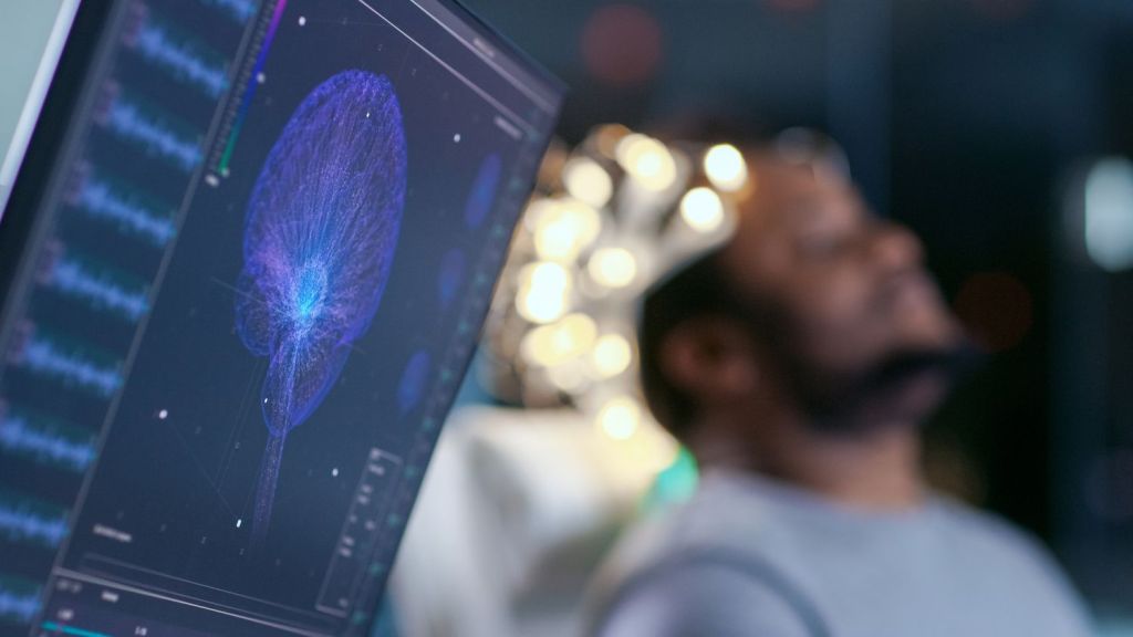
An EEG test records electrical activity in the brain using small electrodes placed on the scalp. Preparation is simple—patients are generally advised to eat and drink normally beforehand. The test is painless and non-invasive.
EEG and Sleep Apnea
Sleep apnea is a common disorder characterized by interrupted breathing during sleep, leading to reduced oxygen supply to the brain. EEG can help detect abnormal brain activity associated with these breathing pauses.
- EEG monitors brainwaves during sleep to identify disruptions caused by apnea events.
- Technicians may ask patients to perform certain activities to stimulate different brain regions during the test.
- Automatic apnea detection uses features like inter-band energy ratios from multi-band EEG data, distinguishing apnea from non-apnea events with classifiers such as K-nearest neighbor.
Research shows that during sleep apnea episodes, spectral power in lower frequency EEG bands increases, indicating heightened cortical arousal. This can negatively affect memory consolidation and working memory performance, as information flow between brain regions is suppressed during apnea events.
EEG and Sleep Paralysis
Sleep paralysis is a temporary inability to move or speak when falling asleep or waking up. It is often associated with REM (rapid eye movement) sleep, vivid dreams, and emotional arousal, and may also be linked to obstructive sleep apnea.
- Symptoms can include hallucinations, such as sensing a presence in the room or pressure on the chest.
- Episodes are usually isolated but can be frightening and occasionally recur, especially in adolescents and young adults.
Improving sleep hygiene—such as maintaining a regular sleep schedule, avoiding caffeine and alcohol, and minimizing light exposure before bedtime—can help reduce sleep paralysis episodes. Keeping a sleep diary may also help identify triggers.
For frequent or chronic symptoms, EEG testing can help rule out underlying causes, such as brain lesions or REM Behavior Disorder (RBD).
EEG and Parasomnia
Parasomnias are abnormal behaviors during sleep, including sleepwalking, night terrors, and nightmares. EEG is often used to diagnose or monitor these conditions and differentiate them from other sleep disorders.
- EEG involves attaching electrodes to the scalp with adhesive or an elastic cap, connected to equipment that records brainwaves.
- The test usually lasts 20–40 minutes, with a neurologist comparing results to normal brainwave patterns.
For parasomnia evaluation, doctors may also recommend improving sleep hygiene and avoiding substances like alcohol or drugs before bedtime. In cases of nocturnal frontal lobe epilepsy, EEG can reveal interictal abnormalities that help distinguish epilepsy from parasomnias.
EEG and Epilepsy
EEG is commonly used to assess and manage epilepsy, especially when seizures occur during sleep. The test helps doctors identify the type and origin of seizures.
- Electrodes are placed on the scalp and connected to a computer that records brain activity.
- Patients must remain still during the test, which may require sleeping while being monitored.
- Video monitoring may be used alongside EEG to observe physical movements.
For example, a patient with nocturnal convulsive episodes may have a normal awake EEG, but a sleep EEG can reveal patterns like centrotemporal spikes, confirming diagnoses such as Rolandic epilepsy. Treatment can then be tailored accordingly.
Frequently Asked Questions
What is an EEG and how is it used for sleep disorders?
An EEG is a test that records the brain’s electrical activity using scalp electrodes. It helps diagnose sleep disorders by identifying abnormal brainwave patterns during different sleep stages.
How does EEG help diagnose sleep apnea?
EEG can detect changes in brainwave activity during apnea events, such as increased spectral power in lower frequency bands, which signal disrupted sleep and cortical arousal.
Can EEG detect causes of sleep paralysis?
Yes, EEG can help rule out underlying neurological causes of sleep paralysis, such as REM Behavior Disorder or brain lesions, especially if episodes are frequent or severe.
Is EEG useful for parasomnia and epilepsy?
EEG is valuable for distinguishing parasomnias from nocturnal epilepsy and for diagnosing epilepsy types that manifest during sleep, guiding appropriate treatment.
How should I prepare for an EEG test?
Eat and drink normally before your EEG. Avoid caffeine or alcohol if advised by your doctor. The test is non-invasive and typically lasts 20–40 minutes.
Are there any risks with EEG testing?
EEG is a safe, non-invasive procedure with no significant risks. Some people may experience mild skin irritation from the electrodes.
For more information about sleep disorders and EEG testing, visit The Sleep Loft.


